Brain Injury
Overview
Mild traumatic brain injury (TBI) affects millions of people each year, from car accident victims to soldiers exposed to IED blast waves. These injuries result in a wide range of symptoms, including concussion, hemorrhage, edema, diffuse axonal injury and vasospasm. The mechanisms of mechanotransduction in TBI are currently poorly understood. We are employing a series of in vitro assays, e.g. high speed stretchers, magnetic tweezers, traction force microscopy and a blast wave bioreactor, to mimic intracranial stresses in TBI and measure their cell-level effects. We aim to elucidate the molecular mechanisms of the injury, in order to develop effective therapeutic solutions.
Neuronal Injury:
Primary Investigators: Matthew Hemphill, Borna Dabiri, Josue Goss
In order to study the mechanisms of mechanotransduction following TBI, we developed a high speed stretcher model which mimics the sudden mechanical stimulus associated with injury. We aim to understand how cells respond to a rapid onset of stress or strain and how this response may initiate the pathological cascade which ultimately leads to TBI. We are currently subjecting populations of neurons cultured on flexible silicon elastomer substrates to a rapid onset stimulus delivered by the high speed stretcher. We identify injured neurons in vitro by distinct morphological alterations which are similar to those associated with diffuse axonal injury (DAI) observed following TBI in vivo. We have recently found that certain cell adhesion structures through which forces are delivered to the neuron may influence the onset of injury.
A 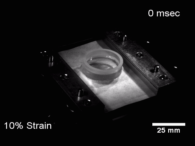
| B 
|
Figure: In vitro model of TBI in a population of neurons
A: | Movie of high speed stretcher |
B: | Example of neurons subjected to stretch (red arrows indicate morphology changes consistent with DAI in vivo |
In order to further elucidate the cellular mechanisms underlying neuronal injury, we employ a technique called Magnetic Tweezers to deliver a local mechanical stimulus to specific regions of the neuron. This technique consists of using an electromagnet to pull on small magnetic beads which are bound to the neuron. By coating the beads with various compounds, we can control the cellular structures through which forces are delivered to the neuron. We have recently found that when we deliver forces through beads coated with fibronectin (FN), an extracellular matrix protein which has binding sites for specific integrin receptors, the extent of injury is much greater than when beads are coated with a substance that does not specifically bind integrins. These results are consistent with the high speed stretcher experiments and suggest that integrin mediated mechanical injury may be a possible explanation of neuronal injury following TBI.
A 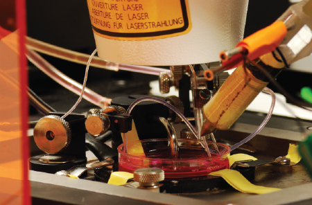
| 
|
Figure: In vitro model of TBI in a single neuron
A: | Magnetic tweezer experimental setup |
B: | Movie of neuron subjected to bead pull (red dot indicates location of bound bead) |
Vascular Injury:
Primary Investigators: Patrick Alford, Ph.D; Sam Felton
Vasospasm of the cerebrovasculature can occur following TBI, especially blast-induced TBI, and is a potentially lethal dysfunction. It is characterized by a chronic vascular hypercontraction followed by cell proliferation, matrix remodeling, and arterial occlusion. While the role of chronically elevated luminal pressures is well established in arterial thickening, the response to rapid, acute pressure increases is not. Therefore, we developed a technique to study this response using the high speed stretcher and vascular smooth muscle MTFs (see the Tissue Engineering section for more on MTFs). We found that when an engineered arterial lamellae was subjected to a single rapid stretch, a strain dependent cellular response was initiated which affected the overall tissue function. We measured changes in vascular contractility and linked these with alterations in expression of contractile phenotypic markers, suggesting that a single mechanical injury may facilitate vascular smooth muscle phenotypic switching which could potentially account for the onset of cerebral vasospasm following TBI.
A 
| B 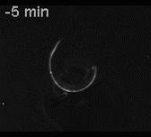
|
Figure: In vitro model of TBI in the vasculature
A: | Engineered arterial lamellae are subjected to a single high speed stretch |
B: | Time lapse movie of induced vascular construct contraction |