Bio-Inspired Nanotextiles
Overview
- A new technique of nanofiber fabrication using the rotary jet spinning for regenerative medicine and other industrial applications
- A mathematical model of rotary jet spinning to predict fiber diameter under different processing conditions
- Manufacturing protein nanoFabrics using extracellular matrix proteins
A new technique of nanofiber fabrication using the rotary jet spinning for regenerative medicine and other industrial applications
Primary Investigator: Mohammad R. Badrossamay, Ph.D
We have developed an effective technique for the generation of continuous fibers and non-woven fabrics with nanometer size fiber diameters by using high-speed mechanical rotation of polymeric solutions through a perforated rotary reservoir. Termed, Rotary jet-spinning (RJS), has several advantages in comparison with other nanofiber fabrication methods: (a) the technique does not require high-voltage electric fields, (b) the apparatus is simple to implement, (c) nanofiber structures can be fabricated into an aligned 3D structure or any arbitrary shape by varying the collector geometry, (d) fiber morphology (beaded, textured, or smooth), fiber diameters, and web porosity can be manipulated by altering the process variables, (e) fiber fabrication is independent of solution conductivity, (f) RJS is easily applicable to polymer emulsions and suspensions, and (g) RJS is capable of substantially higher production rates as compared to standard electrospinning.
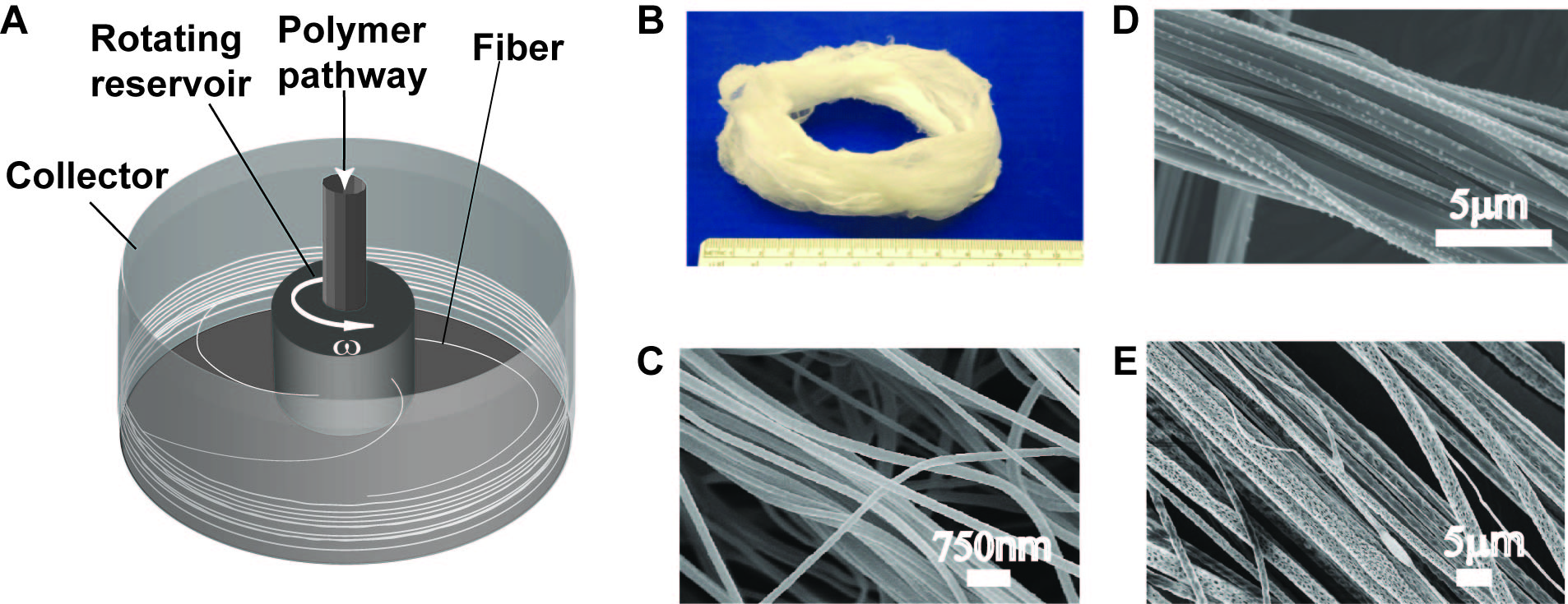
FIGURE: Schematic of rotary jet-spinning (RJS) system
A: Rotary jet spinning is capable of forming 3D structures with any arbitrary shape.
B: Scanning electron micrographs of smooth nanofibers (C), decorated (D) and textured (E).
A mathematical model of rotary jet spinning to predict fiber diameter under different processing conditions
Primary Investigator: Holly McIlwee
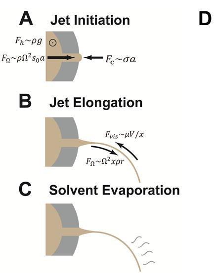
Currently we are studying the formation of nanofibers being produced in our lab via Rotary Jet Spinning from a physical point of view. Using the tools of fluidynamics, we have computed scaling laws that permit us to decrease the diameter and at the same time ensure the continuity of nanofibers, in terms of a few set of tunable laboratory parameters. These scaling laws can be easily translated into phase diagrams for obtaining fibers of desirable characteristics, when design and solution parameters fall into a well defined range.p
| 
|
FIGURE: Three stages of fiber formation in Rotary Jet-Spinning.
A: Jet initiation
B: Jet elongation
C: Solvent evaporation
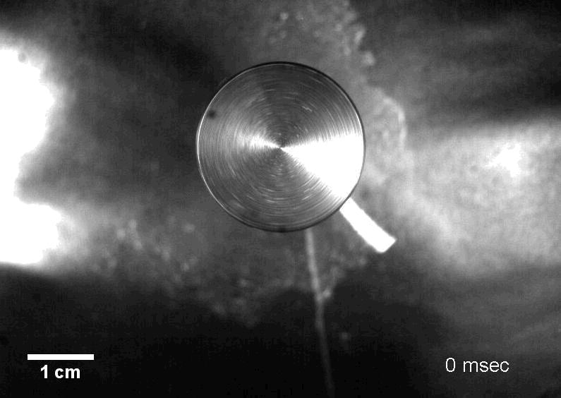
D: Movie shows nanofiber formation by Rotary Jet-Spinning (RJS) captured by high speed camera
Manufacturing protein nanoFabrics using extracellular matrix proteins for applications ranging from biophotonic devices to neuronal tissue engineering
Primary Investigator: Leila Deravi, Ph.D
We have developed a technique for manufacturing bio-inspired nanoFabrics using the model protein Fibronectin (FN). FN nanoFabrics are synthesized using micro-contact printing onto thermosensitive, polymer substrates. At low temperatures (T< 32ºC), the polymer substrate dissolves, and our pre-patterned protein pops off the substrate as free-standing fibrillar networks, or nanoFabrics. When relaxed FN nanoFabrics are mechanically strained, they exhibit a 6-fold extension without failure, likely due to protein extension under strain. By identifying how mechanical strain affects protein conformation within fabrics, we can begin to design a new range of hyper-elastic textiles with similar chemical properties.

FIGURE: Manufacturing FN nanoFabrics.
A: We built FN networks in the form of nanometer thick fabrics by releasing micropatterned FN from a thermosensitive substrate. Scale bar 20 µm.
B: Atomic force micrograph of FN nanoFabrics after release demonstrates nanoscale thickness.
C: Scanning electron micrograph of FN nanoFabrics after release demonstrates millimeter scale dimensions. Scale bar 50 µm.